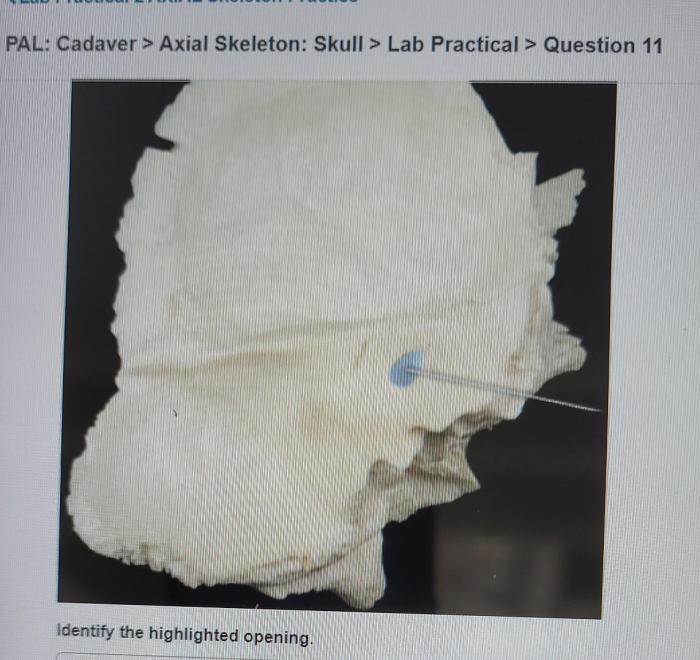
Embarking on pal cadaver axial skeleton skull lab practical question 11, we delve into the intricate realm of human anatomy, meticulously examining the structures of the skull. This practical exploration unveils the significance of these structures, fostering a deeper understanding of their crucial roles in safeguarding and supporting the brain and other vital organs.
The skull, a marvel of anatomical engineering, exhibits a complex morphology, composed of various bones that interlock seamlessly. Sutures and joints facilitate movement, while foramina and canals provide passageways for nerves and blood vessels. Muscles, intricately connected to the skull, orchestrate facial expressions and mastication.
Understanding the clinical significance of the skull underscores its protective role and highlights the potential consequences of skull fractures.
Pal Cadaver Axial Skeleton Skull Lab Practical Question 11

Pal Cadaver Axial Skeleton Skull Lab Practical Question 11 provides an opportunity to examine the anatomy of the human skull firsthand. By observing the various structures, students can gain a deeper understanding of the skull’s morphology and its significance in the study of human anatomy.
The structures observed in this practical session include the frontal bone, parietal bone, temporal bone, occipital bone, sphenoid bone, ethmoid bone, nasal bone, lacrimal bone, zygomatic bone, maxilla, mandible, and hyoid bone. These bones form the protective casing for the brain and other vital structures within the head.
Studying the skull’s anatomy is crucial for understanding the biomechanics of the head and neck region. It helps students comprehend the intricate relationships between the bones, muscles, and other structures in this area.
Skull Anatomy and Morphology
The human skull is a complex structure composed of 22 bones, including 8 cranial bones and 14 facial bones. It can be divided into two main parts: the cranium and the facial skeleton.
The cranium, located in the upper portion of the skull, encloses and protects the brain. It consists of the frontal bone, parietal bones, temporal bones, occipital bone, sphenoid bone, and ethmoid bone. These bones are joined together by immovable joints called sutures.
The facial skeleton forms the lower portion of the skull and is responsible for supporting the facial features. It includes the nasal bones, lacrimal bones, zygomatic bones, maxilla, mandible, and hyoid bone.
Sutures and Joints of the Skull

The bones of the skull are connected by various types of joints, including sutures, syndesmoses, and synchondroses.
Sutures are immovable joints that connect the bones of the cranium. They are formed by the interlocking edges of adjacent bones and are filled with fibrous connective tissue. The most prominent sutures include the coronal suture, sagittal suture, and lambdoid suture.
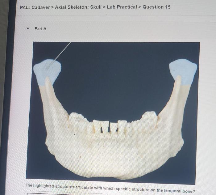
Syndesmoses are slightly movable joints that connect the bones of the facial skeleton. They are formed by a band of fibrous connective tissue that unites the bones. An example of a syndesmosis is the syndesmosis between the mandible and the temporal bone.
Synchondroses are cartilaginous joints that connect the bones of the skull during childhood. They are gradually replaced by bone as the individual matures. An example of a synchondrosis is the synchondrosis between the occipital bone and the sphenoid bone.
Foramina and Canals of the Skull

| Name | Location | Function |
|---|---|---|
| Foramen magnum | Occipital bone | Transmits the spinal cord |
| Jugular foramen | Occipital bone | Transmits the jugular vein, vagus nerve, and glossopharyngeal nerve |
| Hypoglossal canal | Occipital bone | Transmits the hypoglossal nerve |
| Internal acoustic meatus | Temporal bone | Transmits the facial nerve, vestibulocochlear nerve, and blood vessels |
| Carotid canal | Temporal bone | Transmits the internal carotid artery |
| Optic canal | Sphenoid bone | Transmits the optic nerve |
| Superior orbital fissure | Sphenoid bone | Transmits the oculomotor nerve, trochlear nerve, abducens nerve, and ophthalmic nerve |
| Inferior orbital fissure | Maxilla | Transmits the maxillary nerve and infraorbital nerve |
Muscles of the Skull

- Frontalis:Originates from the frontal bone and inserts into the skin of the forehead. Function: Raises the eyebrows.
- Orbicularis oculi:Originates from the medial orbital margin and inserts into the skin around the eye. Function: Closes the eyelids.
- Corrugator supercilii:Originates from the medial orbital margin and inserts into the skin of the eyebrow. Function: Draws the eyebrows together.
- Procerus:Originates from the nasal bone and inserts into the skin of the forehead. Function: Draws the skin of the forehead down.
- Nasalis:Originates from the maxilla and inserts into the skin of the nose. Function: Compresses the nostrils.
- Depressor septi nasi:Originates from the maxilla and inserts into the nasal septum. Function: Lowers the nasal septum.
- Zygomaticus major:Originates from the zygomatic bone and inserts into the skin of the cheek. Function: Raises the corner of the mouth.
- Zygomaticus minor:Originates from the zygomatic bone and inserts into the skin of the cheek. Function: Raises the corner of the mouth.
- Risorius:Originates from the fascia over the masseter muscle and inserts into the skin of the cheek. Function: Draws the corner of the mouth laterally.
- Masseter:Originates from the zygomatic arch and inserts into the mandible. Function: Elevates the mandible.
- Temporalis:Originates from the temporal fossa and inserts into the coronoid process of the mandible. Function: Elevates and retracts the mandible.
- Pterygoid muscles:Originate from the pterygoid plates of the sphenoid bone and insert into the mandible. Function: Medial and lateral pterygoid muscles elevate the mandible, while the lateral pterygoid muscle also protrudes the mandible.
Clinical Significance of the Skull: Pal Cadaver Axial Skeleton Skull Lab Practical Question 11
The skull plays a crucial role in protecting the brain and other vital structures within the head. It provides a rigid framework that shields these delicate tissues from external impacts and injuries.
Skull fractures are common injuries that can result from blunt or penetrating trauma. The severity of a skull fracture depends on the location and extent of the fracture. Some skull fractures can cause significant damage to the brain and other structures, while others may be less severe.
Common types of skull fractures include:
- Linear fractures:These are simple, non-displaced fractures that do not involve the underlying brain tissue.
- Depressed fractures:These fractures occur when a portion of the skull is pushed inward, potentially damaging the underlying brain tissue.
- Basilar skull fractures:These fractures involve the base of the skull and can be particularly dangerous due to their proximity to vital structures such as the brain stem and cranial nerves.
User Queries
What is the significance of the foramina and canals in the skull?
Foramina and canals serve as passageways for nerves and blood vessels, ensuring the proper functioning of the brain and other structures within the skull.
How do the muscles attached to the skull contribute to its function?
These muscles facilitate facial expressions, mastication, and other essential movements, demonstrating the dynamic nature of the skull’s structure.
What are the potential consequences of skull fractures?
Skull fractures can lead to a range of complications, including brain damage, bleeding, and nerve damage, emphasizing the critical protective role of the skull.